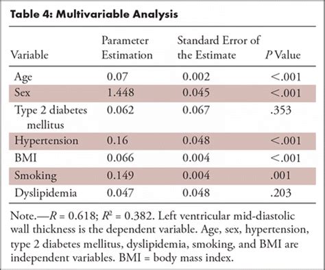
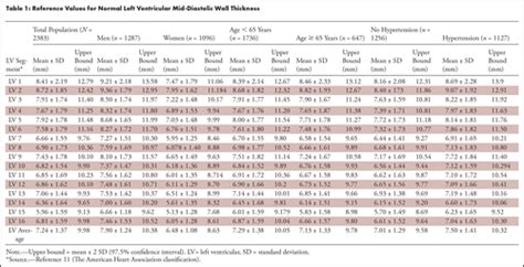
lv myocardium | ivs diastolic thickness normal range lv myocardium Increased left ventricular myocardial thickness (LVMT) is a feature of several cardiac diseases. The purpose of this study was to establish standard reference values of normal LVMT with cardiac magnetic resonance and to assess variation with image acquisition plane, . New Delhi | Updated: November 22, 2022 10:46 IST. Follow Us. One day before the opening ceremony of the 2022 FIFA World Cup, two of football’s biggest players, Cristiano Ronaldo and Lionel Messi, shared a picture that raked up millions of likes within hours of being posted.
0 · normal left ventricular wall thickness
1 · normal left ventricle wall thickness
2 · normal Lv wall thickness
3 · left ventricular non compaction symptoms
4 · ivs diastolic thickness normal range
5 · Lv wall thickness on echo
6 · Lv wall thickness normal values
7 · 17 segments of the heart
Credit24 aizdevums internetā – naudas kredīts tavām iecerēm no 100 līdz 7000 €. Piesakies online un saņem naudu 10 minūtes pēc aizdevuma apstiprināšanas!
Increased left ventricular myocardial thickness (LVMT) is a feature of several cardiac diseases. The purpose of this study was to establish standard reference values of normal LVMT with cardiac magnetic resonance and to assess variation with image acquisition plane, .

PK G´«Xoa«, mimetypeapplication/epub+zipPK G´«X .However, the pathologist’s view of infarcted myocardium lacks insights into the in .In recent years, diagnostic testing to evaluate the presence and extent of .Although excessive trabeculation is present, the presentation of ventricular dilatation, low EF, .
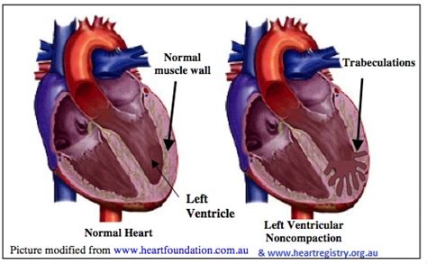
However, the pathologist’s view of infarcted myocardium lacks insights into the .In left ventricular noncompaction (LVNC), there is retarded myocardial morphogenesis and persistence of the trabecular meshwork (B). The ability to acquire LVNC is supported by case reports and studies demonstrating .
This review will focus on clinical manifestations and diagnosis of LVNC as an . In recent years, diagnostic testing to evaluate the presence and extent of viable .
Myocardial ischemia occurs when blood flow to the heart muscle (myocardium) .
Left ventricular (LV) wall thickening or LV hypertrophy (LVH) is commonly . Increased left ventricular myocardial thickness (LVMT) is a feature of several cardiac diseases. The purpose of this study was to establish standard reference values of normal LVMT with cardiac magnetic resonance and to assess variation with image acquisition plane, demographics, and left ventricular function. Overview. Left ventricular hypertrophy Enlarge image. Left ventricular hypertrophy is a thickening of the wall of the heart's main pumping chamber, called the left ventricle. This thickening may increase pressure within the heart. The condition can .
Rapid advances in cardiac computed tomography (CT) have enabled the characterization of left ventricular (LV) myocardial diseases based on LV anatomical morphology, function, density, and enhancement pattern.Although excessive trabeculation is present, the presentation of ventricular dilatation, low EF, and nonischemic myocardial scar and genetic abnormality is the same as in dilated cardiomyopathy. Patient treatment is based on the symptoms and the prognostic risks of arrhythmia, stroke, and contractile impairment. However, the pathologist’s view of infarcted myocardium lacks insights into the in vivo positioning of the LV walls. CMR imaging with delayed contrast enhancement (CE-CMR) has emerged as a new anatomic gold standard technique that provides precise identification of infarcted myocardium in vivo.In left ventricular noncompaction (LVNC), there is retarded myocardial morphogenesis and persistence of the trabecular meshwork (B). The ability to acquire LVNC is supported by case reports and studies demonstrating increased LV trabeculation developing on serial echocardiographic assessment (9–11).
This review will focus on clinical manifestations and diagnosis of LVNC as an isolated disorder distinct from other clinical settings in which non-compacted myocardium may be seen in association with other cardiac and noncardiac abnormalities. In recent years, diagnostic testing to evaluate the presence and extent of viable but dysfunctional myocardium has become an important component of the clinical assessment of patients with chronic CAD and LV dysfunction. Myocardial ischemia occurs when blood flow to the heart muscle (myocardium) is obstructed by a partial or complete blockage of a coronary artery by a buildup of plaques (atherosclerosis). If the plaques rupture, you can have a heart attack (myocardial infarction). Left ventricular (LV) wall thickening or LV hypertrophy (LVH) is commonly observed in clinical practice [1]. Hypertrophic cardiomyopathy (HCM) is the most common genetic cardiomyopathy and is characterized by LVH without an obvious cause [2, 3].
Increased left ventricular myocardial thickness (LVMT) is a feature of several cardiac diseases. The purpose of this study was to establish standard reference values of normal LVMT with cardiac magnetic resonance and to assess variation with image acquisition plane, demographics, and left ventricular function. Overview. Left ventricular hypertrophy Enlarge image. Left ventricular hypertrophy is a thickening of the wall of the heart's main pumping chamber, called the left ventricle. This thickening may increase pressure within the heart. The condition can .
Rapid advances in cardiac computed tomography (CT) have enabled the characterization of left ventricular (LV) myocardial diseases based on LV anatomical morphology, function, density, and enhancement pattern.Although excessive trabeculation is present, the presentation of ventricular dilatation, low EF, and nonischemic myocardial scar and genetic abnormality is the same as in dilated cardiomyopathy. Patient treatment is based on the symptoms and the prognostic risks of arrhythmia, stroke, and contractile impairment. However, the pathologist’s view of infarcted myocardium lacks insights into the in vivo positioning of the LV walls. CMR imaging with delayed contrast enhancement (CE-CMR) has emerged as a new anatomic gold standard technique that provides precise identification of infarcted myocardium in vivo.In left ventricular noncompaction (LVNC), there is retarded myocardial morphogenesis and persistence of the trabecular meshwork (B). The ability to acquire LVNC is supported by case reports and studies demonstrating increased LV trabeculation developing on serial echocardiographic assessment (9–11).

This review will focus on clinical manifestations and diagnosis of LVNC as an isolated disorder distinct from other clinical settings in which non-compacted myocardium may be seen in association with other cardiac and noncardiac abnormalities.
normal left ventricular wall thickness
In recent years, diagnostic testing to evaluate the presence and extent of viable but dysfunctional myocardium has become an important component of the clinical assessment of patients with chronic CAD and LV dysfunction. Myocardial ischemia occurs when blood flow to the heart muscle (myocardium) is obstructed by a partial or complete blockage of a coronary artery by a buildup of plaques (atherosclerosis). If the plaques rupture, you can have a heart attack (myocardial infarction).

prada aoyama

Spec Sheet for ZUMMESH-5A-LV. Download. Add to project. Added to project. Crestron creates world-class commercial lighting control solutions that utilize leading-edge technology for scalable, reliable lighting control. Find an Agent.
lv myocardium|ivs diastolic thickness normal range


