lv motion | Dynamic left ventricular outflow tract obstruction (LVOTO) lv motion LV Volume Measurement 2D Method s LVID Diastole (LVIDD) Inner edge to inner edge, perpendicular to the long axis of the LV, at or immediately below the level of the mitral valve leaflet tips. Perform at end-diastole (defined as the first frame after mitral valve closure or the frame with the largest LV dimensions/volume.) Each The Rolex Air King is made for the man looking for a finely crafted piece with a timeless design and one who appreciates quality and fine craftsmanship. Remaining relatively unchanged throughout its history, the Rolex .
0 · THE AMERICAN SOCIETY OF ECHOCARDIOGRAPHY RECOMMENDATIONS FOR CARDIAC
1 · LV MOTION
2 · Dynamic left ventricular outflow tract obstruction (LVOTO)
DARING AND PANACHE. Rolex presents the new generation of its Oyster .
LV MOTION. Skip to content Search. Language. English. English Español Search. Log in Cart. Item added to your cart View cart. Check out Continue shopping. LV BELT LV BELT Regular .LV Volume Measurement 2D Method s LVID Diastole (LVIDD) Inner edge to inner edge, perpendicular to the long axis of the LV, at or immediately below the level of the mitral valve leaflet tips. Perform at end-diastole (defined as the first frame after mitral valve closure or the frame with the largest LV dimensions/volume.) EachLVOTO is caused by fast-flowing blood through the LV outflow tract which pulls the mitral valve anteriorly (towards the LV outflow tract) due to a Venturi effect. This is known as systolic anterior motion (SAM) of the mitral valve.LV MOTION. Skip to content Search. Language. English. English Español Search. Log in Cart. Item added to your cart View cart. Check out Continue shopping. LV BELT LV BELT Regular price .00 USD Regular price Sale price .00 USD .
Comparison of conventional and contrast-enhanced echocardiography, biplane cine angiography, and cardiac magnetic resonance for the detection of regional wall motion abnormalities. The study underlines the utility of contrast-enhanced echocardiography in comparison to the other methods.Herein we review the conventional assessment of LV systolic function and examine the role of speckle-tracking echocardiography (STE), a new method to assess LV function. We also highlight the role of STE in the assessment and management of cardiac and noncardiac disease, including detection of subclinical LV dysfunction.
Left ventricular outflow tract obstruction (LVOTO) is commonly associated with systolic anterior motion (SAM) of the mitral valve. Congenital heart disease is an important cause in the paediatric population.
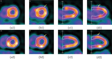
The analysis of left ventricle (LV) wall motion is a critical step for understanding cardiac functioning mechanisms and clinical diagnosis of ventricular diseases. We present a novel approach for 3D motion modeling and analysis of LV wall . Echocardiographic evaluation of wall motion (WM) is a simple, well-validated method to assess segmental left ventricular (LV) function. 1,2 The presence of qualitative WM abnormalities has been demonstrated to be an independent predictor of cardiovascular events in groups of patients with myocardial infarction (MI), 3,4 unstable angina, 5 .Heart motion includes repeated contractions of all four chambers of the heart, with the largest parts being the right ventricle and left ventricle (LV). The range of LV wall motion is normally about 1 cm, yet it can reach up to 3 cm in some regions.Left ventricular (LV) twist is suggested as an important index of contractility and a potential marker of myocardial dysfunction in the diseased heart.
LV Volume Measurement 2D Method s LVID Diastole (LVIDD) Inner edge to inner edge, perpendicular to the long axis of the LV, at or immediately below the level of the mitral valve leaflet tips. Perform at end-diastole (defined as the first frame after mitral valve closure or the frame with the largest LV dimensions/volume.) EachLVOTO is caused by fast-flowing blood through the LV outflow tract which pulls the mitral valve anteriorly (towards the LV outflow tract) due to a Venturi effect. This is known as systolic anterior motion (SAM) of the mitral valve.LV MOTION. Skip to content Search. Language. English. English Español Search. Log in Cart. Item added to your cart View cart. Check out Continue shopping. LV BELT LV BELT Regular price .00 USD Regular price Sale price .00 USD .Comparison of conventional and contrast-enhanced echocardiography, biplane cine angiography, and cardiac magnetic resonance for the detection of regional wall motion abnormalities. The study underlines the utility of contrast-enhanced echocardiography in comparison to the other methods.
Herein we review the conventional assessment of LV systolic function and examine the role of speckle-tracking echocardiography (STE), a new method to assess LV function. We also highlight the role of STE in the assessment and management of cardiac and noncardiac disease, including detection of subclinical LV dysfunction.
Left ventricular outflow tract obstruction (LVOTO) is commonly associated with systolic anterior motion (SAM) of the mitral valve. Congenital heart disease is an important cause in the paediatric population.The analysis of left ventricle (LV) wall motion is a critical step for understanding cardiac functioning mechanisms and clinical diagnosis of ventricular diseases. We present a novel approach for 3D motion modeling and analysis of LV wall . Echocardiographic evaluation of wall motion (WM) is a simple, well-validated method to assess segmental left ventricular (LV) function. 1,2 The presence of qualitative WM abnormalities has been demonstrated to be an independent predictor of cardiovascular events in groups of patients with myocardial infarction (MI), 3,4 unstable angina, 5 .Heart motion includes repeated contractions of all four chambers of the heart, with the largest parts being the right ventricle and left ventricle (LV). The range of LV wall motion is normally about 1 cm, yet it can reach up to 3 cm in some regions.
cost of a birkin bag

THE AMERICAN SOCIETY OF ECHOCARDIOGRAPHY RECOMMENDATIONS FOR CARDIAC
LV MOTION

Rolex Submariner Date Listing: $19,800 Rolex Submariner Two-Tone Stainless .
lv motion|Dynamic left ventricular outflow tract obstruction (LVOTO)


